
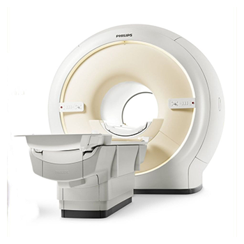
3TESLA MRI
A 3-tesla magnetic field is twice as powerful as the fields used in conventional high-field MRI scanners, and as much as 15 times stronger than low-field or open MRI scanners. This results in a clearer and more complete image. Stronger magnetic fields also produce better images of soft tissues and organs than standard MRI scanners.3T MRI is the fastest diagnostic magnetic resonance imaging technology available. The computer generated images can be sent off-site for immediate analysis. These exams can be up to 100 times faster than standard MRI exams.
Another advantage of 3 Tesla MRI is that the exams offer more comfort than typical MRI exams. Some patients may feel anxious or claustrophobic in traditional MRI scanners. 3 Tesla MRI offers a more spacious bore, or tube, and the patient’s head can remain outside of the bore for scans that don’t involve the spinal cord, neck, or head.
CT Scan 128 Slice Ultrafast
Our Philips ingenuity 128 slice CT is one of the most advanced CT systems in diagnostic radiology field used extensively in neural, cardiac, pulmonary, abdominal, musculoskeletal, trauma and paediatric imaging. This system delivers image quality, dose efficiency and rapid reconstruction.
The system enables faster scan times and lower patient exposure to radiation, while delivering unmatched image quality. 128 slice CT offers a comprehensive range of clinical applications including high resolution Cardiac & Coronary imaging, CT Angiography, 3D Reconstruction and MPR, Virtual Endoscopy, Oncology and Pediatic Imaging. I–DOSE 4 software and rate responsive tool kit has taken image quality of CT CORONARY ANGIOGRAPHY to a amazingly new level even in faster heart rate.

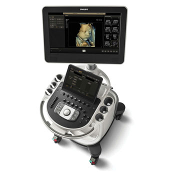
USG & color doppler Affinity 870
Our Philips Affinity 30 and Affinity 50 colour Doppler system is one of the most advanced ultrasound platforms in the world that make it possible to visualize anything accessible to ultrasound. It offers real time 4 D visualization of abdominal, obstetrics, gynaecological, paediatric, superficial structures and vascular imaging. Our very high frequency probe can imagine nerves, nail bed, small tendons and subcutaneous tissues. Trans rectal USG is perfect for imaging of prostate, bladder, rectal and Para rectal lesions. Very high resolution probes and hockey stick probes are specially designed to large small joints, nerves and tendons
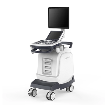
Fibroscan
FibroScan Testing is a recently FDA-approved non-invasive diagnostic device used to measure liver scarring or fibrosis caused by a number of liver diseases. Similar to undergoing a conventional liver ultrasound exam, outpatient FibroScan testing is quick, painless, easy, and provides a non-surgical alternative to the traditional liver biopsy to assess liver damage.In this technique, a 50-MHz wave is passed into the liver from a small transducer on the end of an ultrasound probe . The probe also has a transducer on the end that can measure the velocity of the shear wave (in meters per second) as this wave passes through the liver. The shear wave velocity can then be converted into liver stiffness, which is expressed in kilopascals. Essentially, the technology measures the velocity of the sound wave passing through the liver and then converts that measurement into a liver stiffness measurement; the entire process is often referred to as liver ultrasonographic elastography.
Mammography
We offer Mammography for Breast Cancer detection on our Alleger Venus mammography system, which is one of the most reliable systems available. Breast cancer is one of major risks for women over the age of 40 and a yearly mammogram is the most reliable method of ensuring early diagnosis of this disease, which can be treated effectively, if detected early.

X-Ray
Conventional x rays are still very useful in orthopaedic and other diagnostic field and are gold standard in few cases. Hi-scan has high frequency 500mA x ray unit with CR converter for quality x ray imaging.
Orthoscanogram
Computerized orthoscanogram for whole spine/ whole lower extremity with digital measurements of lengths and angles.

CBCT
CBCT is an imaging technology that allows dentists to evaluate the underlying bone structure, as well as the nerve pathways and surrounding soft tissues. During a CBCT scan, the imaging machine rotates entirely around the patient’s head. In less than a minute, about 150-200 images are captured from a variety of angles and compiled into a single 3D image.
CBCT scans are quick and in most cases, a full mouth scan only takes about 20-40 seconds. When having a CBCT scan taken, you can expect to be seated while an x-ray arm slowly rotates around your head. To ensure your head remains still during the scan, your dentist may have you rest your head against part of the machine and/or use stabilizers in or around your ears to gently hold your head in place. The scan should cause you no discomfort.
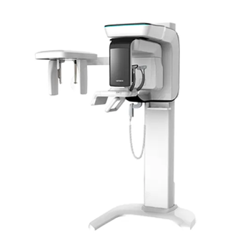
OPG
Updated OPG Machine with Full jaw PAN and TMJ scans, anatomical Programming Radiography (APR), Frontal maxillary sinus scan, center and canine laser centering device and anatomical Programming Radiography (APR).
Lateral cephalometry.

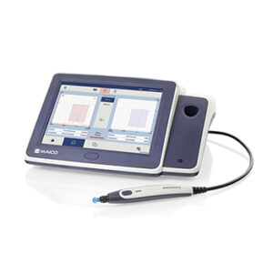
Audiology
- Pure tone audiometry.
- Tympanometry.
- Bera.
- Special test(SISI TDT, ABLB, SDS).
- Hearing aid trails and hearing aids.
- Speech audiometry.
- Speech therapy.
- ETD.
Our Specialities
- 3T advanced MRI (70cm)
- 128 slice CT scan.
- Cardiac CT & CT coronary angiography to facilitate non-invasive diagnosis of cardiac and coronary artery disease which are currently not being done in entire North Bengal and surrounding areas of Bihar, Assam, Bhutan, Nepal & Bangladesh.
- Full range of vascular imaging using CT angiography and high resolution color Doppler imaging.
- High resolution sonography and Doppler, subcutaneous tissues, nerves and joints including nail bed and tendon imaging.
- Fibroscan.
- Audiology solution.
- BMI and body composition analysis which is not being done as a part of routine diagnostic procedures at any other centre.
- Foetal echocardiography using dedicated USG machine and software.
- Trans rectal ultrasonography which is not being used in this part of country.
